Back pain can be frustrating and sometimes alarming, especially when symptoms point to a more serious issue like a herniated disk. An MRI is considered the most accurate imaging test to identify and evaluate herniated disks, providing detailed images that help pinpoint the exact location, size, and severity of the problem. Knowing what to expect from an MRI and how it helps in diagnosing a herniated disk can make the overall process less stressful for anyone seeking answers.
An MRI scan can reveal more than just the presence of a herniated disk—it can show whether it is pressing on nearby nerves and the extent of any spinal damage. This information is vital for doctors to create effective treatment plans and offer precise recommendations for recovery. For those experiencing symptoms like persistent back pain or numbness, understanding how MRI aids in diagnosing a herniated disk can be a crucial step toward relief.
Key Takeaways
- A herniated disk can be accurately identified and evaluated with an MRI.
- MRI results guide both diagnosis and treatment decisions.
- Knowing what an MRI shows helps patients make informed care choices.
Understanding Herniated Disks
A herniated disk occurs when the soft inner core of a spinal disc pushes through its tougher exterior, which can lead to nerve irritation and pain. Several factors contribute to their development, and the symptoms vary based on the location and severity of the herniation.
What Is a Herniated Disk
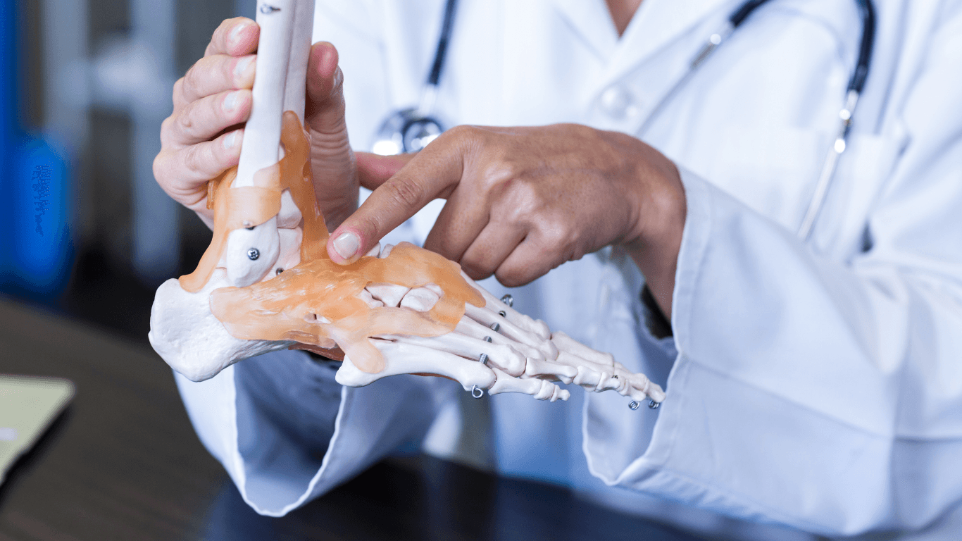
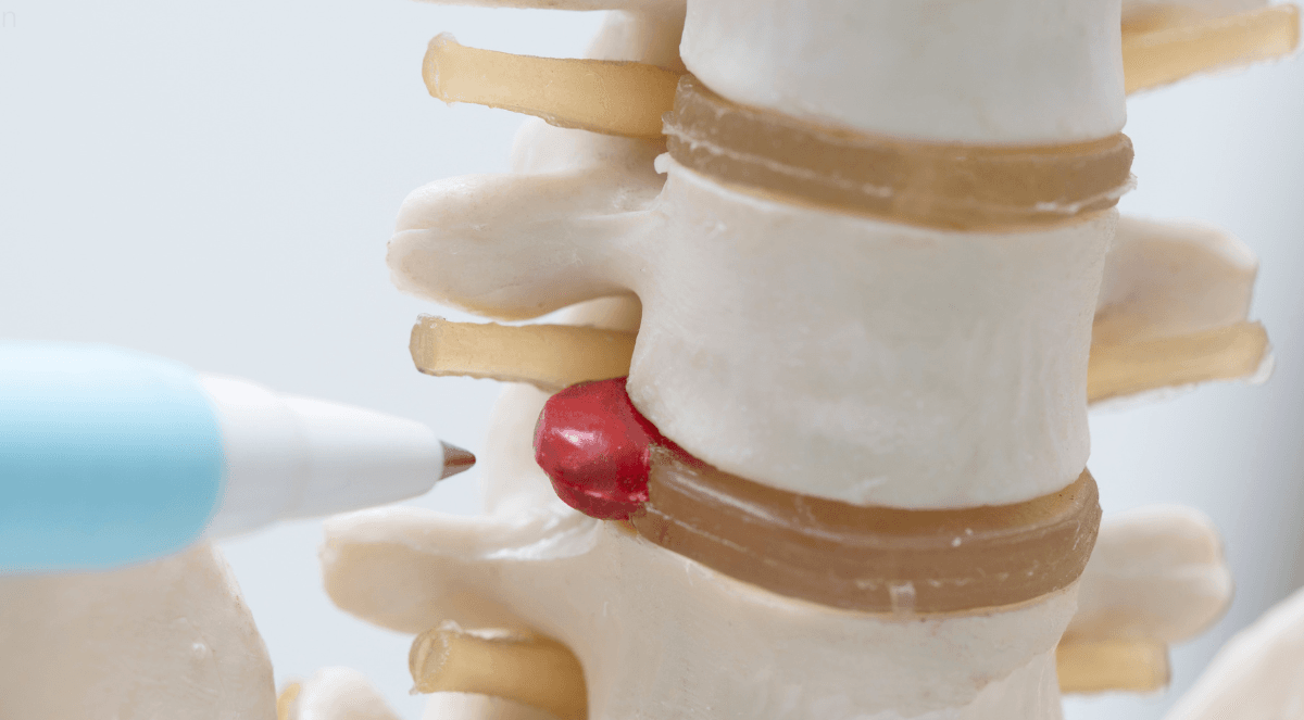
A herniated disk, also known as a slipped or ruptured disc, affects the intervertebral discs that act as cushions between the bones of the spine. Each disc has a soft, gel-like center (nucleus pulposus) surrounded by a tough, fibrous outer layer (annulus fibrosus).
When the outer layer weakens or tears, part of the inner material may protrude out. This bulging or leaking can put pressure on nearby nerves or the spinal cord, causing pain or other neurological symptoms.
Herniations most commonly occur in the lumbar spine (lower back), but they can affect any part of the spine. MRI is the preferred method for clearly identifying herniated discs and visualizing the extent of nerve compression.
Common Causes and Risk Factors
Several factors can cause or increase the risk of a disk herniation:
- Age-related degeneration: Discs lose water content and flexibility with age, making them more prone to tears.
- Sudden trauma: Accidents, falls, or lifting objects incorrectly can cause acute injury.
- Repetitive strain: Jobs or activities involving frequent bending, twisting, or heavy lifting can strain spinal discs.
Other factors include genetics, obesity, a sedentary lifestyle, and smoking. A combination of age-related wear and tear with physical stress is often responsible. Some people may also have congenital conditions that affect spinal disc strength.
Risk is highest for those in their 30s to 50s, but herniations can happen at any age, depending on the specific stressors and lifestyle factors involved.
MRI for Herniated Disk Diagnosis
MRI scans are the leading imaging method for diagnosing herniated disks. They provide detailed images that help physicians determine the exact location, severity, and nature of the disk problem.
How MRI Identifies a Herniated Disk
An MRI uses a combination of magnetic fields and radio waves to generate high-resolution images of spinal structures. When a herniated disk is present, the MRI shows the displacement of the intervertebral disk material and its relationship to adjacent nerves.
Doctors look for bulging, protrusion, or extrusion of the disk’s contents. On the images, a herniated disk typically appears as a deformation or an outpouching of the normally smooth disk margin. Nerve root compression, swelling, and spinal cord involvement can also be seen directly.
Detailed images from MRI scans allow clinicians to assess whether the herniation is pressing on the spinal cord or nerve roots, which is directly linked to symptoms such as pain, numbness, or weakness. MRI can also show degeneration or other spinal abnormalities, providing a comprehensive view of the spine’s condition.
Benefits of MRI Over Other Imaging Methods
MRI is superior to X-rays and CT scans for visualizing soft tissues such as the intervertebral disks and nerves. Unlike X-rays, which only reveal bone structure, MRI images include the disks, nerves, muscles, and spinal cord.
Key benefits of MRI include:
- No radiation exposure
- Clear visualization of soft tissue structures
- Ability to detect nerve impingement or inflammation
- Non-invasive and generally safe for most patients
Doctors are able to pinpoint the exact location and extent of disk herniation with an MRI, allowing for more accurate treatment planning. MRI also helps rule out other causes of back pain by showing nearby anatomy in detail. To understand how MRIs compare in identifying herniated disks, see this discussion on MRI benefits.
MRI Procedure: What to Expect
During an MRI scan for a herniated disk, the patient lies flat on a motorized table that slides into a large, cylindrical scanner. The process is painless and typically lasts between 20 to 45 minutes, depending on the detail required.
The patient will need to remain very still to ensure clear images. Loud thumping or tapping sounds are common, but earplugs or headphones are often provided for comfort. In some cases, a contrast agent may be injected to enhance image clarity, especially if prior surgery or complex anatomy is involved.
Patients are advised to remove all metallic items before the procedure since the magnet can interact with metals. The MRI technologist is present throughout to monitor and assist, and communication with the staff is possible at any time.
Interpreting MRI Results for Herniated Discs
MRI scans offer detailed images that help physicians assess the condition of herniated discs, their impact on surrounding nerves, and the extent of disc displacement or protrusion.
Common MRI Findings
In cases of disc herniation, MRI images often reveal displacement or bulging of the intervertebral disc material. On T1-weighted images, the affected disc usually appears darker, while on T2-weighted images, it may appear brighter due to fluid content. Radiologists examine both sagittal and axial views to detect changes in disc shape and structure.
Typical findings include reduced disc height, irregular disc contours, and evidence of nerve root compression. The proximity and relationship between the herniated disc and adjacent neural structures, such as the spinal cord or nerve roots, are carefully analyzed. Inflammatory changes or nerve root impingement can be inferred from altered fat and fluid patterns in nearby tissues. While herniations can occur anywhere along the spine, they are most frequently observed in the lumbar region, where they commonly contribute to lower back and leg symptoms.
Assessing Severity on MRI
MRI scans help distinguish the severity of herniation, typically classified as mild, moderate, or severe. Mild cases may show slight disc bulging without nerve contact, often referred to as protrusions. Moderate herniations display more pronounced displacement, with partial nerve contact that may result in mild compression or irritation.
Severe herniations are characterized by substantial extrusion of disc material or sequestration—where fragments have broken off and migrated, applying direct pressure on nerve roots. Indicators such as deformation of the spinal canal, compression of nerve roots, and decreased cerebrospinal fluid around nerves suggest higher severity.
Clinicians evaluate these findings by examining the extent of nerve impingement, signal changes in neural tissue, and the overall degree of disc displacement. These insights are essential for determining the appropriate treatment plan, including whether surgical intervention may be necessary.
Frequently Asked Questions
MRI scans provide detailed images to identify herniated discs, assess severity, and guide treatment decisions. The scan helps differentiate between various disc abnormalities and pinpoints the affected spinal level.
What are the symptoms indicating an MRI is needed for diagnosing disc herniation?
Signs that may prompt a doctor to recommend an MRI include persistent back pain, numbness, weakness, or tingling in the legs or feet. Loss of bladder or bowel control and severe pain radiating down the leg can also be indications. MRI imaging is necessary to confirm disc herniation.
How can an MRI differentiate between a bulging disc and a herniated disc?
On MRI, a bulging disc shows a broad-based, symmetrical outpouching of the disc, but the outer layer remains intact. A herniated disc appears as focal displacement of the inner disc material that has broken through the outer layer, often compressing nearby nerves. These details can be identified on high-resolution MRI imaging.
What does a severe herniated disc look like on an MRI?
A severe herniated disc typically appears as a large mass of disc material that has moved out of its normal location and is compressing the spinal cord or nerve roots. The affected area may show significant nerve impingement, displacement, or even loss of spinal fluid space on the scan. Images will clearly indicate nerve compression or crowding.
Can an MRI accurately reveal all stages of disc herniation?
MRI can detect various stages of disc disease, from minor bulges to extensive herniation. Early changes like mild bulging are often visible, but the test is most effective at showing moderate to severe herniation, nerve compression, and associated swelling or inflammation. Advanced imaging can also identify secondary effects such as nerve root irritation or displacement.